PETVET clinics are well-equipped, full-service, small animal veterinary practices providing comprehensive diagnostic, medical, surgical and dental care. We aim to offer quality service, giving your pet the maximum opportunity to lead a long and happy life.
PETVET clinics are well-equipped, full-service, small animal veterinary practices providing comprehensive diagnostic, medical, surgical and dental care. We aim to offer quality service, giving your pet the maximum opportunity to lead a long and happy life.
ALL THE CARE YOUR PET NEEDS!
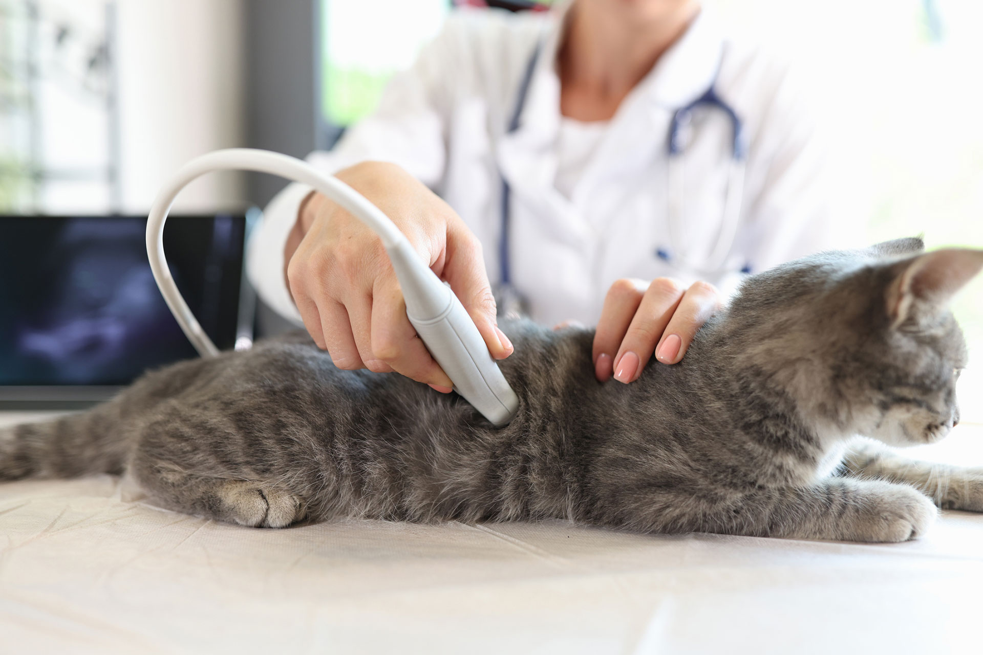
An Ultrasound scan, most commonly associated with pregnancy, is also a useful tool we can use to help diagnose or stage many different diseases in your pet.
What is Ultrasound?
Ultrasound is a non-invasive procedure similar to an x-ray. It works by sending very high frequency sound waves through body tissues and recording the waves as they are reflected back. Sophisticated computer programmes in the Ultrasound machine transform those reflections into detailed images of the internal organs and other objects. As the sound waves are transmitted continuously an ultrasound scan produces a moving picture of an organ or body part as it is actually functioning. The pattern of reflected sound waves can also be processed to generate images and sounds of blood flow through the heart and other organs.
Most organs except those containing air such as the lungs can be evaluated. Commonly the heart, liver, pancreas, kidneys, intestines, spleen, urinary bladder and other organs located in the abdomen are evaluated. Eyes, tendons, muscles, bones and joints as well as the brain in very young animals can also be imaged. The benefits of ultrasound are enormous and are used for a wide range of condition:-
- Diseases that are difficult to diagnose
- Diseases undetected without invasive surgery
- Diseases that otherwise would require sophisticated and expensive diagnostic procedures
- Tissue biopsies can be taken by using an ultrasound scan to guide the biopsy needle to the required area through a tiny incision in the skin.
- Abnormal fluid accumulation in the chest cavity, around the heart or within the abdominal cavity is easily removed under ultrasound guidance.
- An ultrasound may confirm that surgery is essential e.g. to remove a foreign object in the bowel
- An ultrasound may identify conditions that surgery will not help such as multiple invasive tumours
Although there are other parts of the body that can be studied with ultrasound, abdominal and cardiac ultrasound are the most common in veterinary medicine.
Abdominal Ultrasound
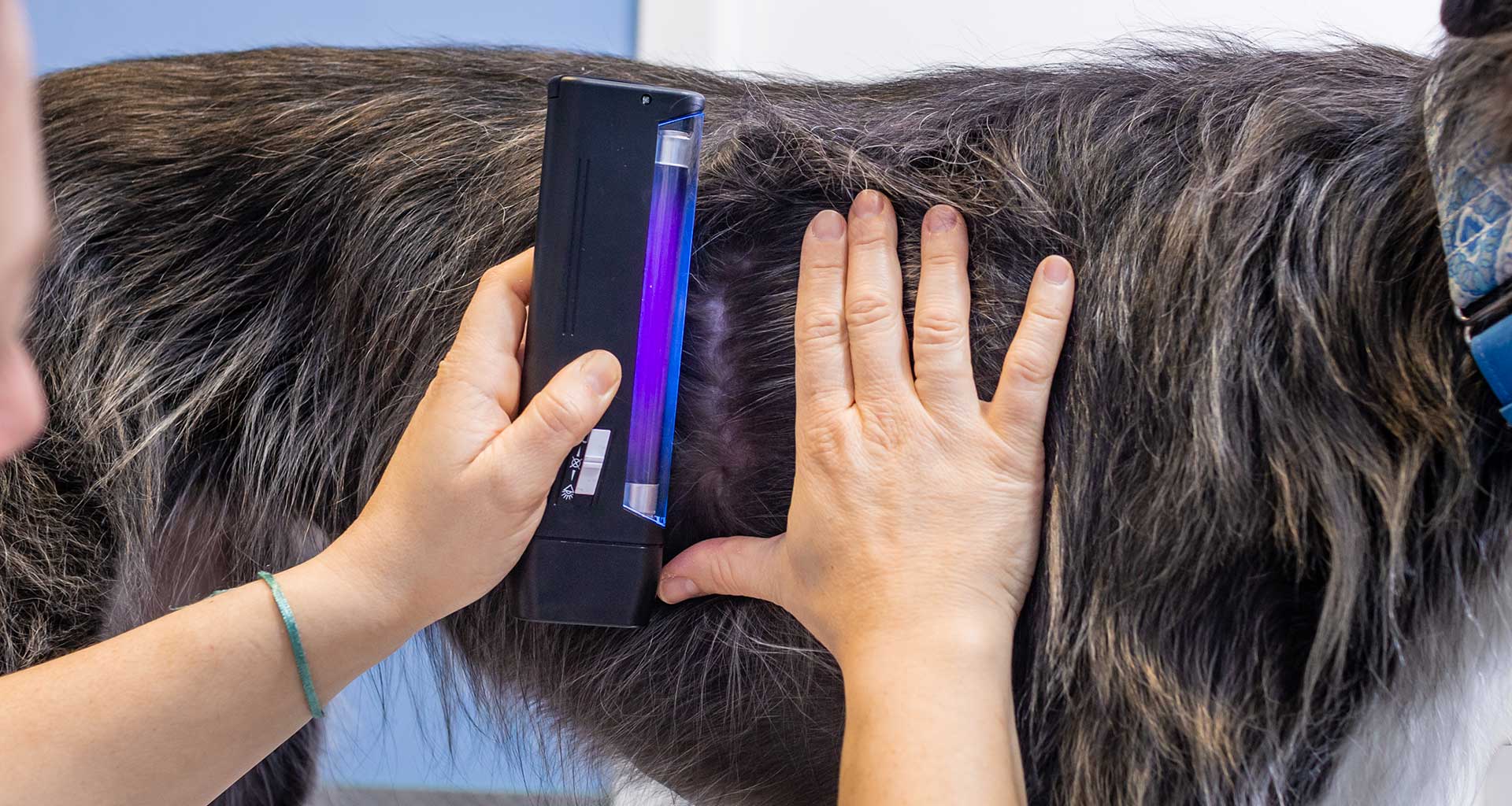
All Ultrasound examinations require the probe to closely contact the skin. This enables the sound waves to be efficiently transmitted and the echoes detected. To produce the best possible picture, the hair must be shaved from the abdomen. After the hair is shaved a layer of gel is used to provide a good acoustic coupling between the skin and the probe. The probe is scanned across the skin of the abdomen to examine the organs or regions of interest. Most pets require sedation for the exam to be completed. Anaesthesia will normally be required for more painful procedures such as ultrasound guided biop- sies.
Abdominal ultrasound is used to evaluate pets with symptoms such as vomiting, diarrhoea, straining to urinate, urinating blood and to screen for cancers. It can also be helpful in cases of reproductive abnormalities, weight loss and to detect early pregnancy. When physical examination and blood tests indicate a problem with a particular organ, an ultrasonic examination can provide additional information or even a diagnosis.
Heart Ultrasound (Echocardiography)
An ultrasound scan of the heart is known as an echocardiogram. The procedure is very similar to abdominal ultrasound. Hair is clipped from the chest behind the elbow to ensure a good quality study. Sedation is sometimes needed to relax the patient enough to lie still on the examination table.
Cardiac ultrasound allows us ‘to see inside your pet’s heart’. We can assess the function of the heart valves, the thickness of the heart muscle, contractions of the heart and the flow of blood through the heart chambers and over the valves. Fluid buildup around the heart (pericardial effusion) and growths around the heart can also be identified.
Ultrasound has become a very useful diagnostic tool in veterinary medicine. To a large extent, ultrasound has replaced ex- ploratory abdominal surgery. Along with radiography, ultrasound can also be used to diagnose and treat most heart diseases that occur in dogs and cats.
Preparing for Ultrasound
In order to obtain a high quality diagnostic image most ultrasounds require the hair to be clipped over the area to be scanned. The ultrasound probe must contact the skin closely without any air pockets. It is a good idea to give your dog a bath a day or two before their ultrasound to reduce dirt in the coat. This is not a good idea for cats but a good brush will help.
Preparing for Sedation or Anaesthesia
Sedation or low dose pain killers and relaxants are more common than anaesthesia for Ultrasounds unless they are combined with other procedures such as X-rays or Surgery. All anaesthetics involve some risk. For most ultrasound patients this risk is very small but inadequate preparation can increase these risks.
Some patients are very sensitive to sedative and anaesthetic drugs. The most common reaction is to vomit, either while they are going under or when they are waking up. Because anaesthetic drugs reduce the gag and swallow reflexes they may inhale vomited material. Vomited material inhaled into the lungs can lead to severe pneumonia. It is important than all food is with- held from 9.00pm the night before admission to allow the stomach to empty. Remember that patients who go outside at night might scavenge if they are hungry. This applies particularly to cats so it is best to confine cats overnight.
As sedatives and anaesthetics can have some adverse effects on organ function, particularly the kidneys, it is important that your pet does not become dehydrated. Fresh drinking water should be available to your pet during the night before ultra- sonography. For most procedures your pet will receive an intravenous drip or other fluid support.
Pre-anaesthetic Blood Tests
Some patients may have underlying problems that can be made worse by anaesthesia. Often they are older patients, but some breeds are prone to congenital problems, such as kidney disease or bleeding disorders even when they are young. Blood tests before ultrasound can screen for potential problems and allow us to modify the procedure accordingly.
On the Day of the Ultrasound
If your pet is in hospital we will take care of their pre-anaesthetic preparation and medication for you.
Admission is between 7.30am and 8.30am. We will double check the procedure, your pets preparation and check if there is anything else to be done while admitted. If you would like a pre anaesthetic blood screen now is a good time to request it.
If your pet requires anaesthesia for their procedure we will administer a pre operative sedative and painkiller. If your pet is either very nervous or aggressive with strangers it is very helpful to let us know so that we can give this to your pet in your presence. Once the sedative has calmed your pet they will usually accept handling by our hospital staff.
Your pets’ blankets, bed or toys should be left at home. They can become a focus for anxiety and end up soiled, lost in the wash, or may be mutilated. Occasionally they may be eaten, causing an obstruction in the bowel, and creating a need for more surgery. Your pet will be provided with comfortable bedding and will appreciate their own when they return home. We will also provide a collar and lead for your dog while it is in hospital. They may be removed and replaced a number of times during their stay in hospital and as a result, your own lead can easily become misplaced.
Please note, results are generally not available on the day of the procedure. Please arrange a revisit or telemedicine consultation with your referring vet to discuss the results.
Check List Before an Ultrasound
- Access to water overnight
- Skin clean
- Medication sorted
- Admission 7.30am-8.30am
- No Bedding or Toys please
- We will provide a collar and lead for dogs
- Cats confined to a secure carry cage